
Celiac Disease Testing - CAM 315
Description
Celiac disease is a hereditary, chronic autoimmune disorder triggered by the ingestion of gluten, a protein found in wheat, rye, and barley. When an individual with celiac disease ingests gluten, the body mounts an immune response that attacks the small intestine. These attacks lead to damage on the villi within the small intestine, inhibiting nutrient absorption1
Regulatory Status
Many labs have developed specific tests that they must validate and perform in house. These laboratory-developed tests (LDTs) are regulated by the Centers for Medicare and Medicaid (CMS) as high-complexity tests under the Clinical Laboratory Improvement Amendments of 1988 (CLIA ’88). LDTs are not approved or cleared by the U. S. Food and Drug Administration; however, FDA clearance or approval is not currently required for clinical use.
The Quanta Lite Celiac Screen ELISA test for tissue transglutaminase/gliadin and the Quanta Lite Celiac DGP Screen by Inova Diagnostics, Inc. were approved by the FDA on 01/28/1999 and 12/13/2006, respectively. Quanta Plex Celiac IgA and IgG profiles by Inova Diagnostics, Inc. were approved on 03/14/2007 and 06/20/2007.
EliA Celikey IgG for use with the EliA Celikey IgG Immunoassay by Phadia US, Inc. was approved by the FDA on 12/26/2006.
The FIDIS Celiac on the FIDS Analyser and FIDIS CELIAC kit by Biomedical Diagnostics S.A. were approved by the FDA on 09/24/2004 and 03/29/2006, respectively.
The IMMULISA CELIAC ELISA testing systems for gliadin IgA/IgG and TTG IgA/IgG by IMMCO Diagnostics, Inc. were approved on 02/04/2010 and 03/10/2010. IMMCO’s IMMULISA enhanced celiac fusion (TTG/DGP) IgA/IgG antibody ELISA system was approved on 10/25/2013.
Bio-Rad Laboratories’ Bioplex 2200 Celiac IgA IgG kits were approved on 09/19/2013. The IgX Plex Celiac qualitative assay and Ig Plex Celiac DG panel by SQI diagnostics systems, Inc. were approved by the FDA on 06/02/2011 and 11/06/2014, respectively.
SQI Diagnostics received FDA clearance for the Ig plex Celiac DGP which detects IgA and IgG antibodies to deamidated gliadin peptide (DGP) and tissue transglutaminase (tTG) in human serum. This was approved by the FDA on Nov 06, 2014.40
Inova Diagnostics received FDA clearance on June 16, 2021, for the Aptiva Celiac Disease IgA Reagent, which is an “immunoassay utilizing particle-based multi-analyte technology for the semi-quantitative determination of anti-tissue transglutaminase IgA autoantibodies and anti-deamidated gliadin peptide IgA antibodies in human serum.” They also received approval for an IgG Reagent in 2021. It can be used to diagnose celiac disease and dermatitis herpetiformis.41
Policy:
Application of coverage criteria is dependent upon an individual’s benefit coverage at the time of the request.
- For individuals who have been diagnosed with celiac disease and who are IgA sufficient, serologic testing with IgA anti-tissue transglutaminase (TTG) is considered MEDICALLY NECESSARY at the following intervals:
- At the first follow-up visit 3 – 6 months after diagnosis.
- Every 6 months until normalization of anti-TTG levels has occurred.
- Every 12 – 24 months thereafter.
- For individuals who have been diagnosed with celiac disease and who are IgA deficient, testing for IgG endomysial antibodies, IgG deamidated gliadin peptide, or IgG TTG is considered MEDICALLY NECESSARY at the following intervals:
- At the first follow-up visit 3 – 6 months after diagnosis.
- Every 6 months until normalization of IgG levels has occurred.
- Every 12 – 24 months thereafter.
- For individuals with signs and symptoms of celiac disease (see Note 1), serologic testing with the IgA anti-TTG and the total IgA test for the diagnosis of celiac disease is considered MEDICALLY NECESSARY.
- For individuals at risk for celiac disease (see Note 1), when IgA anti-TTG is negative or weakly positive, testing for IgA endomysial antibodies is considered MEDICALLY NECESSARY.
- For individuals with clinical suspicion of celiac disease (see Note 1) with an IgA deficiency, testing for IgG endomysial antibodies, IgG deamidated gliadin peptide, or IgG TTG is considered MEDICALLY NECESSARY.
- Testing for IgA and IgG antibodies to deamidated gliadin peptides is considered MEDICALLY NECESSARY in any of the following situations:
- For individuals under 2 years of age with a clinical suspicion of celiac disease (see Note 1).
- For individuals over 2 years of age as a substitute for anti-TTG testing.
- For confirmation of celiac disease in individuals at high risk for celiac disease, regardless of the result of celiac disease serology testing, pathological examination of tissue obtained from a biopsy of the small intestine is considered MEDICALLY NECESSARY.
- Rapid antigen point-of-care testing for anti-TTG is considered NOT MEDICALLY NECESSARY.
- Panel testing, multiplex testing, or multi-analyte testing (for more than two analytes) for the diagnosis or evaluation of celiac disease is considered NOT MEDICALLY NECESSARY.
- For asymptomatic individuals not at an increased risk for developing celiac disease (see Note 1), testing for celiac disease is considered NOT MEDICALLY NECESSARY.
The following does not meet coverage criteria due to a lack of available published scientific literature confirming that the test(s) is/are required and beneficial for the diagnosis and treatment of an individual’s illness.
- For the diagnosis of celiac disease, testing for anti-reticulin antibodies is considered NOT MEDICALLY NECESSARY.
- For the evaluation of celiac disease , testing of stool or saliva samples is considered NOT MEDICALLY NECESSARY.
- Serologic testing using an HLA-DQ-gluten tetramer-based assay, including flow cytometry-based HLA-DQ-gluten tetramer assays, is considered NOT MEDICALLY NECESSARY.
NOTES:
Note 1: Signs and symptoms of celiac disease may include, but are not limited to, the following: unexplained chronic or intermittent diarrhea; unexplained weight loss; unexplained chronic or intermittent abdominal pain or bloating; recurrent nausea or vomiting; unexplained iron deficiency anemia; unexplained vitamin B12 or folate deficiency; unexplained liver transaminase elevations; autoimmune hepatitis; dermatitis herpetiformis; Type 1 diabetes; intestinal blockages; unexplained subfertility or miscarriage; unexplained osteoporosis, osteomalacia, or low bone density; and/or primary biliary cirrhosis. Individuals with Down syndrome, Turner syndrome, or Williams-Beuren syndrome are also at high risk for celiac disease. Additionally, in pediatric patients, fatty stools, delayed puberty, amenorrhea, failure to thrive, stunted growth, and/or short stature may also be associated with celiac disease2-4
Table of Terminology
| Term |
Definition |
| AAFP |
American Academy of Family Physicians |
| ACG |
American College of Gastroenterology |
| AEA |
Anti-endomysium antibodies |
| AGA |
American Gastroenterological Association |
| ALT |
Alanine transaminase |
| ANG |
Anti-native gliadin antibodies |
| BSG |
British Society of Gastroenterology |
| BSPGHAN |
British Society of Pediatrics Gastroenterology, Hepatology, and Nutrition |
| CD |
Celiac disease |
| CI |
Confidence interval |
| CLIA ’88 |
Clinical Laboratory Improvement Amendments of 1988 |
| CMS |
Centers for Medicare and Medicaid Services |
| DGP |
Deamidated gliadin peptides |
| DH |
Dermatitis Herpetiformis |
| DTC |
Direct to consumer |
| EGD |
Esophagogastroduodenoscopy |
| ELISA |
Enzyme-linked immunosorbent assay |
| EMA |
Endomysial antibodies |
| ESPGHAN |
European Society for Pediatric Gastroenterology, Hepatology, and Nutrition |
| ESsCD |
European Society for the Study of Coeliac Disease |
| FABP |
Fatty acid-binding protein |
| FDA |
Food and Drug Administration |
| GFD |
Gluten-free diet |
| GI |
Gastrointestinal |
| HBV |
Hepatitis B virus |
| HLA |
Human leukocyte antigen |
| HLA-DQA1 |
Major histocompatibility complex, class II, DQ alpha 1 gene |
| HLA-DQB1 |
Major histocompatibility complex, class II, DQ beta 1 gene |
| HLA-DQ2 |
HLA class II histocompatibility antigen, DQ2 |
| HLA-DQ8 |
HLA class II histocompatibility antigen, DQ8 |
| IgA |
Immunoglobulin A |
| IgG |
Immunoglobulin G |
| LCD |
Local Coverage Determinations |
| LDTs |
Laboratory-developed tests |
| MLPA |
Multiplex ligation-dependent probe amplification |
| NASPGHAN |
North American Society for Pediatric Gastroenterology, Hepatology, and Nutrition |
| NASSCD |
North American Society for the Study of Celiac Disease |
| NCD |
National Coverage Determinations |
| NCGS |
Non celiac gluten sensitivity |
| NGS |
Next-generation sequencing |
| NICE |
National Institute for Health and Care Excellence |
| NIDDK |
National Institute of Diabetes and Digestive and Kidney Diseases |
| PCR |
Polymerase chain reaction |
| PPV |
Positive predictive value |
| RIA |
Radioimmunoassay |
| sIgE |
Specific immunoglobulin-E |
| SSOP |
Sequence-specific oligonucleotide probe |
| T1D |
Type 1 diabetes |
| TIDM |
Type I diabetes mellitus |
| TG2 |
Transglutaminase type 2 |
| TGA-IgA |
IgA-antibodies against transglutaminase 2 |
| TSH |
Thyroid stimulating hormone |
| TTG |
Tissue transglutaminase |
| ULN |
Upper limit of normal |
| USPSTF |
United States Preventative Services Task Force |
| WA |
Wheat allergy |
| WGO |
World Gastroenterology Organization |
| ZED1227 |
Oral transglutaminase 2 inhibitor |
Rationale
Celiac disease (CD) is an autoimmune disease which occurs due to the body’s unfavorable response after the ingestion of gluten. In particular, the body’s immune system attacks the small intestine, leading to damage and inhibiting nutrient absorption.1 The clinical presentation of CD is varied and age dependent. In children, failure to thrive, malnutrition, diarrhea, abdominal pain, and distension are common. In adults, abdominal pain, diarrhea or constipation, bloating, and excessive gas are frequent symptoms. Other gastrointestinal symptoms include unexpected weight loss and distension.5 A high prevalence of CD cases are often found in first degree relatives of CD patients, highlighting genetic aspects of the disease.6 Currently, the only treatment for CD is to maintain a gluten-free diet to ameliorate symptoms and improve the quality of life.7
The National Institute of Diabetes and Digestive and Kidney Diseases provides the following statistics for CD:
- About two million Americans have CD
- About 1% of people worldwide have CD
- The majority of people are undiagnosed
- CD can affect all races, but is present at a higher rate in Caucasians
- CD can affect both genders
- CD is more common among people with chromosomal disorders like Down syndrome, Turner syndrome, and Williams syndrome
- Patients with CD are at risk for Addison’s disease, Hashimoto’s disease, selective IgA deficiency, primary biliary cholangitis, and type 1 diabetes8
CD has a strong genetic component. The two primary genetic factors for CD susceptibility are the human leukocyte antigen (HLA)-DQ2 and DQ8 alleles.9 These genes highlight the role of T cells and the immune response in CD.10 Approximately 90-95% of CD patients have the HLA-DQ2 protein encoded by the HLA-DQA1*05 and DQB1*02 alleles. The remaining CD patients have mutations in the HLA-DQ8 protein encoded by the HLA-DQA1*03 and DQB1*03:02 alleles. Stankovic, et al. (2014) noted that the absence of susceptible HLA-DQ genotypes makes CD “very unlikely, close to 100%.” However, the use of genotyping in diagnosing CD is not without controversy. Paul, et al. (2017) report that 25-40% of white Caucasians are positive for the HLA-DQ2/DQ8 haplotype but that only 0.1-1% of the population will develop CD. They also note that the European guidelines released in 2012 recommend genotyping for HLA-DQ2/DQ8 in children with very high anti-TTG titers, but the authors recommend that “HLA-DQ2/DQ8 testing must not be done to 'screen' or 'diagnose' children” with CD.12
Antibodies for the assessment of CD generally fall into one of two categories: autoantibodies (tTG-IgA, anti-endomysial antibody [EMA-IgA]) or antibodies targeting gliadin (DGP-IgA or IgG, antigliadin antibody (AGA)-IgA or IgG). Endomysial antibodies bind to tissue transglutaminase and produce a characteristic staining pattern. Similarly, anti-endomysial antibodies bind to tTG-2, another tissue transglutaminase. The other category of celiac antibodies involve gliadin, which is a component of gluten. Traditional antigliadin antibody tests (AGA-IgA, AGA-IgG) yielded a false positive rate of up to 20%, so they have been replaced with a deamidated gliadin peptide (DGP).5
Genetic testing for HLA DQ2 and DQ8 may also be used to confirm a CD diagnosis. Serologic and histologic HLA-DQ testing requires the patient to be on a gluten-containing diet, which can be a disadvantage to testing. Recently, testing methods for HLA-DQ-gluten tetramer-based assays using flow-cytometry have been developed; these tests can accurately determine whether the patient is on a gluten-containing or gluten-free diet. The assay has a reported 97% sensitivity and 95% specificity for patients on a gluten-free diet as compared to controls (patients without CD).13 The authors conclude, “This test would allow individuals with suspected celiac disease to avoid gluten challenge and duodenal biopsy, but requires validation in a larger study.”13
Point of care tests, such as the Simtomax®, have been developed, which detects IgA and IgG antibodies against deamidated gliadin peptides (DGP) and provides a response in just ten minutes. The assay can be obtained through a finger prick technique or a plasma sample.14 There are also direct-to-consumer (DTC) tests for CD. The FDA-approved 23andme panel includes CD. This test detects a single nucleotide polymorphism in HLA-DQA1.15
Clinical Utility and Validity
Olen, et al. (2012) evaluated the diagnostic performance and actual costs in clinical practice of immunoglobulin (Ig)G/IgA DGP (deamidated gliadin peptide antibodies) as a complement to IgA-TTG for the diagnosis of pediatric CD. The authors identified 278 children with CD that received a duodenal biopsy. Sensitivity and specificity for tTG were 94% and 86% respectively, but corresponding values for DGP were 91% and 26%. Positive predictive values were 88% for tTG and 51% for DGP. The authors concluded that for diagnosing CD, tTG is superior to DGP, even in children younger than 2 years. Further, combining tTG and DGP does not provide a better trade-off between number of missed cases of CD, number of unnecessary duodenal biopsies, and cost than utilizing tTG alone.16
Sakly, et al. (2012) evaluated the usefulness of anti-DGP antibodies (a-DGP) in the diagnosis of CD. The study included 103 untreated CD patients of all ages and 36 CD patients under a gluten-free diet. The specificity of a-DGP was 93.6% for IgG and 92% for IgA as compared to the 100% for each by anti-endomysium antibodies (AEA) and tTG. The authors concluded that the findings of this study showed “that a-DGP increases neither the sensitivity nor the specificity of AEA and AtTG [anti-tissue transglutaminase antibodies].”17
Bufler, et al. (2015) evaluated the diagnostic performance of three serological tests for CD. A total of 91 children with CD contributed 411 sera samples and were compared to 98 healthy controls. Transglutaminase type 2 (TG2)-IgA, deamidated gliadin peptide (DGP)-IgG, and DGP-IgA were measured. The sensitivity for diagnosis was high for TG2-IgA and DGP-IgG (>90%) but lower for DGP-IgA. Specificity was >97% for all three. Non-adherence to a gluten-free diet was best indicated by positive TG2-IgA. The authors concluded that “combined testing for TG2-IgA and DGP-IgG does not increase the detection rate of CD in IgA competent children compared to TG2-IgA only.”18
Silvester, et al. (2017) performed a meta-analysis to evaluate the “sensitivity and specificity of tTG IgA and EMA [endomysial antibody] IgA assays in identifying patients with celiac disease who have persistent villous atrophy despite a gluten-free diet (GFD).” The authors identified 26 studies for inclusion. The assays were found to have high specificity for identifying patients with persistent villous atrophy (0.83 for tTG IgA, 0.91 for EMA IgA, but with low sensitivity (0.50 for tTG IgA, 0.45 for EMA IgA). No significant difference was seen between pediatric and adult patients. The authors concluded that “we need more-accurate non-invasive markers of mucosal damage in children and adults with celiac disease who are following a GFD.”19
A report by Selleski, et al. (2018) shows that only some of the DQ2/DQ8 alleles were significantly different between pediatric CD patients and pediatric non-CD patients. A total of 97% of the CD patients were positive for at least either DQ2 or DQ8; however, 29.9% of the non-CD patients were also positive for DQ2. In fact, “No significant association was found between DQ2.2 variant and celiac disease in the studied population.”20 Previously, high regard had been given to DQ2.2 variant as being a predisposing variant for CD.21 Finally, a rapid nucleic acid amplification test using multiplex ligation-dependent probe amplification (MLPA) to detect HLA-DQ2.2, HLA-DQ2.5, and HLA-DQ8 has been developed with a reported 100% specificity for those particular genotypes,22 but this test has not been FDA-approved for use in the United States.
Bajor, et al. (2019) performed a meta-analysis focusing on the association between the HLA-DQB1*02 gene doses and the characteristics of CD. The authors identified 24 studies for inclusion in the review and observed that homozygosity of the DQB1*02 allele led to more frequent classical CD (odds ratio [OR] 1.758). The gene dosing effect was more prominent in children (OR: 2.082). Atrophic histology (Marsh grade 3) was more prevalent with a double dose compared to a zero dose (OR: 2.626). No gene dosing effect was seen with diarrhea, age at diagnosis, severity of villous atrophy, or type 1 diabetes. The authors concluded that “A double dose of HLA-DQB1*02 gene seems to predispose patients to developing classical CD and villous atrophy. Risk stratification by HLA-DQB1*02 gene dose requires further clarification due to the limited available evidence.”23
Tangermann, et al. (2019) completed a prospective study which included 1055 patients all tested for CD with the Simtomax point of care test. The Simtomax detects IgA and IgG antibodies against deamidated gliadin peptides (DGP). All results were compared to the gold standard: histologic analysis of duodenal biopsies. Of all patients who participated in this study, the overall CD prevalence was identified at 4.1%; the Simtomax identified CD with a 79% sensitivity, 94% specificity, 37% positive predictive value, and 99% negative predictive value.24 When the adult (n=888) and pediatric (n=167) patients were analyzed separately, the Simtomax was found to identify CD with 100% sensitivity and 95% specificity in adults, and 72% sensitivity in children; the authors note that the Simtomax test detected CD with a lower sensitivity than expected.24
Profaizer, et al. (2020) conducted a study to “evaluate the feasibility of using NGS-based [next-generation sequencing] HLA-B and DQ genotyping for clinical HLA disease association testing and provide direct comparison with the currently used clinical tests, including SSOP [sequence-specific oligonucleotide probe] genotyping, and real-time PCR [polymerase chain reaction] with melting chain analysis.” The researchers focused on HLA alleles related to celiac disease, ankylosing spondylitis, abacavir hypersensitivity, carbamazepine hypersensitivity, and allopurinol hypersensitivity. With regards to CD and from 24 samples tested, there was a discrepancy with the DQB1*03:40 allele with SSOP, real-time PCR, and NGS, but overall, with the different HLA-correlations the data has shown “HLA typing by NGS is superior to the existing clinical methods for identifying HLA alleles associated with disease or drug hypersensitivity and offers a viable approach for high volume clinical diagnostic laboratories,” continuing to demonstrate the clinical utility of NGS and HLA-testing for CD.25
Gould, et al. (2021) evaluated CD serologic testing in asymptomatic patients with type 1 diabetes using immunoglobulin an anti-tissue transglutaminase, as there is an increased risk of type 1 diabetes among CD and vice versa. From screening 2,353 patients, the assay with IgA anti-tissue transglutaminase had a positive predictive value of 85.9% when referenced upper limit of normal and had a sensitivity and specificity of 100% and 38%, respectively. This study indicated the need for thresholds for diagnostic evaluation to be population-specific (i.e. to type 1 diabetics), and not taken from the overall population due to the increased risk.26
Schuppan et al. (2021) assessed the efficacy and safety of a 6-weel treatment with ZED1227, a selective oral transglutaminase 2 inhibitor, at three dose levels compared with placebo, in adults with well-controlled celiac disease who underwent a daily gluten challenge. Their primary endpoint was the attenuation of gluten-induced mucosal damage, measured by the ratio of villus height to crypt death. For this study, 41 patients were assigned to the 10-mg ZED1227 group, 41 patients were assigned to the 100-mg group, and 40 patients were assigned to the placebo group. Each had adequate duodenal-biopsy samples for the assessment of the overall endpoint. “The estimated difference from placebo in the change in the mean ratio of villus height to crypt depth from baseline to week 6 was 0.44 (95% confidence interval [CI], 0.15 to 0.73) in the 10-mg group (P = 0.001), 0.49 (95% CI, 0.20 to 0.77) in the 50-mg group (P<0.001), and 0.48 (95% CI, 0.20 to 0.77) in the 100-mg group (P<0.001). The estimated differences from placebo in the change in intraepithelial lymphocyte density were -2.7 cells per 100 epithelial cells (95% CI, -7.6 to 2.2) in the 10-mg group, -4.2 cells per 100 epithelial cells (95% CI, -8.9 to 0.6) in the 50-mg group, and -9.6 cells per 100 epithelial cells (95% CI, -14.4 to -4.8) in the 100-mg group.” The authors concluded that treatment with ZED1227 attenuated gluten-induced duodenal mucosal damage in patients with celiac disease (Schuppan et al., 2021).
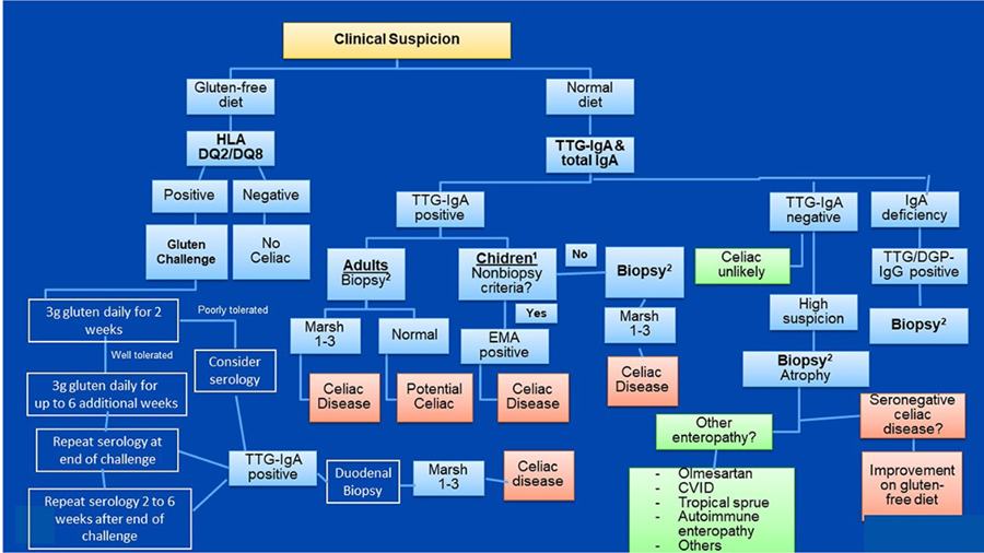
A 2023 update from ACG focused on the diagnosis and management of celiac disease expands upon their previous guidelines. The following are ACG recommendations:
“1A. We [ACG] recommend EGD with multiple duodenal biopsies for confirmation of diagnosis in both children and adults with suspicion of CD (strong recommendation, moderate quality of evidence; dissent 1).
1B. We suggest a combination of high-level TTG IgA (>10× upper limit of normal) with a positive endomysial antibody (EMA) in a second blood sample as reliable tests for diagnosis of CD in children. In symptomatic adults unwilling or unable to undergo upper GI endoscopy, the same criteria may be considered after the fact, as a diagnosis of likely CD (conditional recommendation, moderate quality of evidence; dissent 0).”
ACG explains the above recommendations by citing key concepts:
“1. Multiple biopsies of the duodenum (1 or 2 from bulb and 4 from distal duodenum) are necessary for diagnosis of CD.
2. EGD and duodenal biopsies can also be useful for the differential diagnosis of other malabsorptive disorders or enteropathies.
3. Lymphocytic duodenosis (≥25 intraepithelial lymphocytes per 100 epithelial cells) in the absence of villous atrophy is not specific for CD, and other causes should be considered.”27
Moreover, in the case of screening procedures, ACG states that
“7A. We recommend case finding to increase detection of CD in clinical practice (strong recommendation, low quality of evidence; dissent 0).
7B. We recommend against mass screening for CD in the community (strong recommendation, low quality of evidence; dissent 0),” on the basis that:
“1. Patients with symptoms, signs, or laboratory evidence suggestive of malabsorption, such as chronic diarrhea with weight loss, steatorrhea, abdominal pain, and bloating, should be tested for CD.
2. Patients with symptoms, signs, or laboratory evidence for which CD is a treatable cause should be considered for testing for CD.
3. Patients with a first-degree family member who has a confirmed diagnosis of CD should be tested whether they show possible signs or symptoms or laboratory evidence of CD.
4. Consider testing of asymptomatic relatives with a first-degree family member who has a confirmed diagnosis of CD.”27
Although they note that there is controversy surrounding the best serology approach for children younger than 2 years, ACG notes:
“8A. We recommend the immunoglobulin IgA anti-TTG antibody (TTG-IgA) as the preferred single test for the detection of CD in children younger than 2 years who are not IgA-deficient (strong recommendation, moderate quality of evidence; dissent 0).
8B. We recommend that testing for CD in children with IgA deficiency be performed using IgG-based antibodies (DGP-IgG or TTG-IgG) (strong recommendation; moderate quality of evidence; dissent 0)” because
“1. TTG-IgA and EMA-IgA are reported to be less accurate in children younger than 2 years.
2. Current guidelines recommend that testing for CD in children younger than 2 years include both TTG-IgA and DGP-IgG.”27
Additionally, ACG recommends individuals with type I diabetes mellitus “should be tested for CD if there are any suggestive symptoms or signs. There is evidence that CD is substantially more common in patients with type I diabetes than in the general White population. The estimates vary between 3% and 10%. In children, it has been suggested that yearly or every-other-year screening for CD be undertaken using serology. Patients with type I diabetes who are undergoing upper endoscopy should undergo duodenal biopsies to rule out CD if previous CD testing has not been undertaken.”
Regarding monitoring and follow-up biopsies, the guideline states: “upper endoscopy with intestinal biopsies is recommended for monitoring in cases with lack of clinical response or relapse of symptoms despite a GFD.” A “Follow-up biopsy could be considered for assessment of mucosal healing in adults in the absence of symptoms after 2 years of starting a GFD after shared decision-making between patient and provider.”
With regards to HLA genetic testing, “genetic testing for CD-compatible human leukocyte antigen (HLA) haplotype is not required for diagnosis in all cases but may be helpful in selected situations such as in the context of serology-histology discrepancy. If negative, CD is ruled out. HLA testing is also central to the approach to CD testing for individuals who have already started a GFD before evaluation; in the presence of a CD-compatible haplotype, a gluten challenge can be offered.”27
Lastly, the 2023 ACG recommendation comments on “emerging evidence” which suggests that a non-biopsy diagnosis may be available and accurate with a variety of commercial serology kits and pretest probabilities. However, they do not go so far as to endorse these tests, as “solid information about nonbiopsy diagnosis of CD in the United States is not available yet.”27
Celiac Disease Diagnostic Testing Algorithm.27
American Gastroenterological Association (AGA)
Relative to ongoing monitoring of individuals with celiac disease, the AGA recommends periodic serologic testing.
The AGA published an update on CD testing in 2019. Their new “best practice advice” is as follows:
- “Best Practice Advice 1: Serology is a crucial component of the detection and diagnosis of CD, particularly tissue transglutaminase–immunoglobulin A (TG2-IgA), IgA testing, and less frequently, endomysial IgA testing.”
- “Best Practice Advice 2: Thorough histological analysis of duodenal biopsies with Marsh classification, counting of lymphocytes per high-power field, and morphometry is important for diagnosis as well as for differential diagnosis.”
- “Best Practice Advice 2a: TG2-IgA, at high levels (> ×10 upper normal limit) is a reliable and accurate test for diagnosing active CD. When such a strongly positive TG2-IgA is combined with a positive endomysial antibody in a second blood sample, the positive predictive value for CD is virtually 100%. In adults, esophagogastroduodenoscopy (EGD) and duodenal biopsies may then be performed for purposes of differential diagnosis.”
- “Best Practice Advice 3: IgA deficiency is an infrequent but important explanation for why patients with CD may be negative on IgA isotype testing despite strong suspicion. Measuring total IgA levels, IgG deamidated gliadin antibody tests, and TG2-IgG testing in that circumstance is recommended.”
- “Best Practice Advice 4: IgG isotype testing for TG2 antibody is not specific in the absence of IgA deficiency.”
- “Best Practice Advice 5: In patients found to have CD first by intestinal biopsies, celiac-specific serology should be undertaken as a confirmatory test before initiation of a gluten-free diet (GFD).”
- “Best Practice Advice 6: In patients in whom CD is strongly suspected in the face of negative biopsies, TG2-IgA should still be performed and, if positive, repeat biopsies might be considered either at that time or sometime in the future.”
- “Best Practice Advice 7: Reduction or avoidance of gluten before diagnostic testing is discouraged, as it may reduce the sensitivity of both serology and biopsy testing.”
- “Best Practice Advice 8: When patients have already started on a GFD before diagnosis, we suggest that the patient go back on a normal diet with 3 slices of wheat bread daily preferably for 1 to 3 months before repeat determination of TG2-IgA.”
- “Best Practice Advice 9: Determination of HLA-DQ2/DQ8 has a limited role in the diagnosis of CD. Its value is largely related to its negative predictive value to rule out CD in patients who are seronegative in the face of histologic changes, in patients who did not have serologic confirmation at the time of diagnosis, and in those patients with a historic diagnosis of CD; especially as very young children before the introduction of celiac-specific serology.”28
The AGA’s best advice statements for evaluating refractory celiac disease is recorded below.
“Best Practice Advice 1
In patients believed to have celiac disease who have persistent or recurrent symptoms or signs, the initial diagnosis of celiac disease should be confirmed by review of prior diagnostic testing, including serologies, endoscopies, and histologic findings.
Best Practice Advice 2
In patients with confirmed celiac disease with persistent or recurrent symptoms or signs (nonresponsive celiac disease), ongoing gluten ingestion should be excluded as a cause of these symptoms with serologic testing, dietitian review, and detection of immunogenic peptides in stool or urine. Esophagogastroduodenoscopy with small bowel biopsies should be performed to look for villous atrophy. If villous atrophy persists or the initial diagnosis of celiac disease was not confirmed, consider other causes of villous atrophy, including common variable immunodeficiency, autoimmune enteropathy, tropical sprue, and medication-induced enteropathy.
Best Practice Advice 3
For patients with nonresponsive celiac disease, after exclusion of gluten ingestion, perform a systematic evaluation for other potential causes of symptoms, including functional bowel disorders, microscopic colitis, pancreatic insufficiency, inflammatory bowel disease, lactose or fructose intolerance, and small intestinal bacterial overgrowth.
Best Practice Advice 4
Use flow cytometry, immunohistochemistry, and T-cell receptor rearrangement studies to distinguish between subtypes of refractory celiac disease and to exclude enteropathy-associated T-cell lymphoma. Type 1 refractory celiac disease is characterized by a normal intraepithelial lymphocyte population and type 2 is defined by the presence of an aberrant, clonal intraepithelial lymphocyte population. Consultation with an expert hematopathologist is necessary to interpret these studies.
Best Practice Advice 5
Perform small bowel imaging with capsule endoscopy and computed tomography or magnetic resonance enterography to exclude enteropathy-associated T-cell lymphoma and ulcerative jejunoileitis at initial diagnosis of type 2 refractory celiac disease.
Best Practice Advice 6
Complete a detailed nutritional assessment with investigation of micronutrient and macronutrient deficiencies in patients diagnosed with refractory celiac disease. Check albumin as an independent prognostic factor.”29
European Society for Pediatric Gastroenterology, Hepatology, and Nutrition (ESPGHAN)
Updated and expanded evidence-based guidelines for diagnosing CD were published in 2020 by the ESPGHAN. The following recommendations were included.2
- “We recommend considering testing for CD in children and adolescents with symptoms, signs and conditions shown in Table 2”
- Signs and symptoms in Table 2 include:
- “Gastrointestinal: chronic or intermittent diarrhea, chronic constipation not responding to usual treatment, chronic abdominal pain, distended abdomen, recurrent nausea, recurrent vomiting
- Extraintestinal symptoms: weight loss, failure to thrive, stunted growth/short stature, delayed puberty, amenorrhea, irritability, chronic fatigue, neuropathy, arthritis/arthralgia, chronic iron-deficiency anemia, decreased bone materialization (osteopenia/osteoporosis), repetitive fractures, recurrent aphthous stomatitis, dermatitis herpetiformis-type rash, dental enamel defects, abnormal liver biochemistry
- Specific conditions: first degree relatives with CD, autoimmune conditions: TIDM, thyroid disease, liver disease, Down syndrome, Turner syndrome, Williams-Beuren syndrome, IgA deficiency”
- Signs and symptoms in Table 2 include:
- “HLA- typing does not add to the certainty of the diagnosis if the other criteria for CD diagnosis are fulfilled. Testing for HLA DQ2 and DQ8 may be useful in other circumstances. If no risk alleles are found, CD is unlikely. We recommend that HLA typing is not required in patients with positive TGA-IgA, if they qualify for CD diagnosis with biopsies or if they have high serum TGA-IgA (≥10× ULN) and EMA-IgA positivity. If a patient tests negative for HLA DQ2 and DQ8, the risk of CD is very low, while a positive result does not confirm the diagnosis”
- “Recent studies suggest that the no-biopsy approach to diagnose CD can be applied in asymptomatic children. In asymptomatic children, however, the PPV of high TGA-IgA ≥10× ULN may be lower than in symptomatic children, which needs to be considered during the decision-making process. We give a conditional recommendation that, taking available evidence into account, CD can be diagnosed without duodenal biopsies in asymptomatic children, using the same criteria as in patients with symptoms. We recommend that the decision whether or not to perform diagnostic duodenal biopsies should be made during a shared decision-making process together with the parent(s) and, if appropriate, with the child”
- “The three specific coeliac antibodies (TGA-IgA, EMA-IgA, DGP-IgG) show different performance. TGA-IgA scored highest by a comparison of assay accuracy and is therefore regarded as the most appropriate primary test for CD in the diagnostic work up of children with suspected CD. We recommend that in subjects with normal serum IgA values for age, TGA-IgA should be used as the initial test regardless of age”
- “We recommend testing for total IgA and TGA-IgA as initial screening in children with suspected CD. In patients with low total IgA concentrations, an IgG-based test (DGP, EMA, or TGA) should be performed as a second step. Testing for EMA, DGP or AGA antibodies (IgG and IgA) as initial screening in clinical practice is not recommended”
- “We recommend that for CD diagnosis without biopsies, TGA-IgA serum concentration of at least 10× ULN should be obligatory. Only antibody tests with proper calibrator curve-based calculation and having the 10× ULN value within their measurement range, should be used. We recommend against omitting biopsies in IgA-deficient cases with positive IgG-based serological tests”
- “We recommend that in children with TGA ≥10X ULN, and parents/patient agreement to the no-biopsy approach, the CD diagnosis should be confirmed by a positive EMA-IgA test in a second blood sample”
- “At least 4 biopsies from the distal duodenum and at least 1 from the duodenal bulb should be taken for histology assessment during a gluten-containing diet. Reading of biopsies should be performed on optimally orientated biopsies. A villous to crypt ratio of <2 indicates mucosal lesions. In cases of discordant results between TGA-IgA results and histopathology, re-cutting of biopsies and/or second opinion from an experienced pathologist should be requested.”2
A 2022 position paper on the management and follow-up of children and adolescents with celiac disease stated that:
“3.1 The first follow-up visit should be scheduled 3–6 months after CD diagnosis, but with easy access to the celiac service if earlier advice is needed, and sooner review if there are concerns regarding how the family is coping with the diet, if there are ongoing issues with growth or persistent symptoms or a need to repeat bloodwork earlier. Subsequent visits should be every 6 months until normalization of TGA levels, and every 12–24 months thereafter.”
“3.2. During follow-up patients should be evaluated for:
3.2.I. Gastrointestinal and extraintestinal signs and symptoms.
3.2.II. Anthropometric measurements and growth parameters.
3.2.III. IgA-TGA using the same assay as at diagnosis as a surrogate marker for improvement/healing of the small-bowel mucosa. IgG based tests and RIA based IgA-TGA measurements are not suitable for follow-up in IgA sufficient patients. IgA insufficient patients with CD should be followed with IgG based tests.
3.2.IV. A complete blood cell count, micronutritional status (e.g., hemoglobin, iron, vitamin B12, and vitamin D levels) and ALT measurements, should be performed after clinical evaluation at time of diagnosis. Any abnormality should be followed and deficiencies corrected until normalization. If abnormalities persist, additional diagnoses should be considered and appropriately investigated.
3.2.V. Screening for thyroid disease with TSH and thyroxine (and autoantibodies if indicated) may be considered during follow-up after clinical evaluation at the discretion of the clinician.
3.2.VI. Routine bone-density screening is not recommended.
3.2.VII. HBV antibody levels may be measured in previously immunized patients if this is considered important in the population. A booster dose should be given if inadequate levels are present.”30
A few specific issues of note that may come up during follow-up and management include:
“6.1. How to approach persistent high serum levels of antibodies against tissue-transglutaminase (TGA)?
Lack of decreasing IgA-TGA levels after 6–12 months on a GFD or persisting positive IgA-TGA levels should be assessed by carefully reviewing dietary compliance and testing IgA-TGA using the same test from the same manufacturer.
6.2. When is it necessary to (re)biopsy?
Routine assessment of mucosal healing by small-bowel biopsies is not recommended in children with CD following a GFD. We recommend considering (re) biopsy only in selected CD cases; based on specific clinical grounds, for example, when doubts about the original diagnosis or suspicion of occurrence of an additional condition.
6.3. Refractory celiac disease in children: does it exist?
We recommend properly investigating other causes of an apparent “refractory CD” in children, including ongoing inadvertent ingestion of gluten and other possible concomitant enteropathies, such as Crohn’s disease, autoimmune enteropathy, small-bowel bacterial overgrowth, cow’s milk protein allergy and pancreatic insufficiency.”30
ESPGHAN suggests that “In cases of uncertain CD diagnosis, HLA typing should be performed before gluten-challenge to detect children in whom the occurrence of CD is unlikely.” Moreover, they “recommend the same frequency and follow-up tests in children with CD and T1D as in children with isolated CD, with (additional) special attention to test for thyroid involvement and diabetic retinopathy” and that “developing the follow-up plan in conjunction with an endocrinologist/diabetologist and a dietitian, also considering the need for psychological and social support.”30
North American Society for Pediatric Gastroenterology, Hepatology, and Nutrition (NASPGHAN)
NASPGHAN updated their recommendations in 2015 (published in 2016) for gluten-related disorders, including CD, wheat allergy (WA), and nonceliac gluten sensitivity (NCGS). Concerning who should be tested for gluten-related disorders, “Children with symptoms consistent with gluten-related disorders, or who have self-identified relief of symptoms when avoiding gluten, should undergo testing for CD and/or WA before the elimination of dietary gluten. CD should be an early consideration in those with typical gastrointestinal symptoms such as chronic diarrhea, abdominal pain, distension, and weight loss.” The table below outlines their recommendations for considering CD testing:
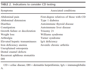
“Children belonging to groups known to be at increased risk for CD may initially have no symptoms, or very minor symptoms, despite having intestinal histologic changes that are characteristic for CD. Included in these groups are first-degree relatives of an index case, people with trisomy 21, Turner syndrome, Williams syndrome, and IgA deficiency, and those with other autoimmune conditions.”31
For initial testing, they recommend the TTG-IgA antibody test due to its reliability and cost-effectiveness. They note that co-testing for serum IgA can be performed to “identify those who have selective IgA deficiency”; however, “use of a panel of antibodies instead of a single tTG-IgA test is not recommended. Although this approach may be associated with a marginal increase in the sensitivity of the test, it decreases the specificity and significantly increases the costs.”31 Testing for serum antibodies against gliadin is less sensitive, reliable, and specific as compared to TTG and EMA.
They do not recommend genetic testing for HLA variants as an initial diagnostic test or screening for CD since up to 40% of the general population contains one of the variant alleles. “Testing for HLA-DQ2/8 is best reserved for patients in whom there is a diagnostic dilemma, such as when there is a discrepancy between the serological and histologic findings or when a GFD [gluten-free diet] has been started before any testing.”31
They do not recommend the use of rapid, point-of-care tests for TTG since these tests do not allow for the quantitative analysis of the antibody.
National Institute for Health and Care Excellence (NICE)
In 2022, NICE published guidance on diagnosing CD. These guidelines state that serological testing should be offered to “people with any of the following: persistent unexplained abdominal or gastrointestinal symptoms, faltering growth, prolonged fatigue, unexpected weight loss, severe or persistent mouth ulcers, unexplained iron, vitamin B12 or folate deficiency, type 1 diabetes, at diagnosis, autoimmune thyroid diseases, at diagnosis, irritable bowel syndrome (in adults), [and] first-degree relatives of people with celiac disease.”3
“Any test is accurate only if a gluten-free containing diet is eaten during the diagnostic process and advise the person not to start a gluten-free diet until diagnosis is confirmed by a specialist, even if the results of a serological test are positive.”3
Further, serological testing for CD could be considered in patients with “metabolic bone disorder (reduced bone mineral density or osteomalacia), unexplained neurological symptoms (particularly peripheral neuropathy or ataxia), unexplained subfertility or miscarriage, persistently raised liver enzymes with unknown cause, dental enamel defects, Down’s syndrome, and Turner syndrome.”3
Finally, regarding serological testing:
- “Test for total IgA and IgA tTG as the first choice
- Use IgA EMA if IgA tTG is weakly positive
- Consider using IgG EMA, IgG DGP or IgG tTG if IgA is deficient (IgA deficiency is defined as total IgA less than 0.07 g per litre).”3
In 2015, the National Institute for Health and Care Excellence (NICE) recommended CD serologic testing in symptomatic young people and adults with the following algorithm:32
- First test for total serum IgA and TTG
- Next test for IgA endomysial antibodies (EMA) if TTG is inconclusive (i.e., weakly positive)
- “Consider using IgG EMA, IgG deamidated gliadin peptide (DGP) or IgG tTG if IgA is deficient”
For children with suspected CD, they recommend:
- First test for total serum IgA and TTG
- “Consider using IgG EMA, IgG DGP or IgG tTG if IgA is deficient”
NICE also recommends offer CD testing for people with any of the following:
- Autoimmune thyroid disease
- Persistent unexplained abdominal or gastrointestinal symptoms
- Irritable bowel syndrome
- Type 1 diabetes
- First-degree relatives (parents, siblings, or children) with coeliac disease
- Other symptoms indicative of possible CD, including faltering growth in children, prolonged fatigue, unexpected weight loss, severe or persistent mouth ulcers, unexplained dietary deficiencies
NICE also recommends considering CD testing for people with the following:
- Metabolic bone disorder
- Unexplained neurological symptoms
- Unexplained subfertility or recurrent miscarriage
- Down’s syndrome or Turner’s syndrome
- Dental enamel defects
- Persistent elevated hepatic enzymes of unknown etiology
They do note that “People who are following a normal diet (containing gluten) should be advised to eat gluten in more than 1 meal every day for at least 6 weeks before testing for coeliac disease.”33
NICE indicates that HLA testing should not be done as part of the initial testing. Also, “Only consider using HLA DQ2 (DQ2.2 and DQ2.5)/DQ8 testing in the diagnosis of coeliac disease in specialist settings (for example, in children who are not having a biopsy, or in people who already have limited gluten ingestion and choose not to have a gluten challenge).”32
United States Preventive Services Task Force (USPSTF)
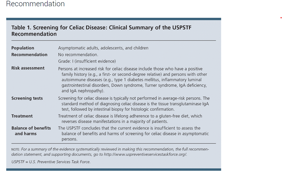
The United States Preventative Services Task Force recently published guidelines on the screening of asymptomatic populations for celiac disease and found that:
“The USPSTF concludes that the current evidence is insufficient to assess the balance of benefits and harms of screening for celiac disease in asymptomatic persons. Evidence is lacking, and the balance of benefits and harms cannot be determined.” However, it was noted that: “Persons at increased risk for celiac disease include those who have a positive family history (eg, a first- or second-degree relative), with an estimated prevalence of 5% to 20%, and persons with other autoimmune diseases (eg, type 1 diabetes mellitus, inflammatory luminal gastrointestinal disorders, Down syndrome, Turner syndrome, IgA deficiency, and IgA nephropathy). Several specialty societies recommend screening in these populations.”34
World Gastroenterology Organisation (WGO) Global Guidelines
The WGO published guidelines on CD testing in 2017. A cascade with “resource-sensitive” options is listed.
The “Gold Standard” lists the following items for diagnosis of CD:
- Celiac disease–specific antibodies: assessment and intestinal biopsy
- Anti-tTG IgA or anti-EMA IgA, and total IgA to exclude IgA deficiency
- In case of selective IgA deficiency, IgG-based tests should be used: anti-DGP, anti-tTG, or EMA (the latter 2 are highly sensitive, but with lower specificity)
- Symptomatic patients with a positive serological test or a titer just below the cut-off (borderline) should be referred for endoscopy with multiple duodenal biopsies to confirm or exclude the diagnosis of celiac disease. Pitfalls in histologic diagnosis are common, and findings are characteristic, but not specific
- Asymptomatic patients with a positive serological test should be retested after consuming a gluten-containing diet for 3 months, to confirm persistent seropositivity before referral for endoscopy
The following items are listed for management of CD:
- Follow-up monitoring, including antibody tests (anti-tTG IgA or DGP-IgG in case of IgA deficiency): after 3 to 6 months in the first year and once a year thereafter in stable patients responding to the gluten-free diet
The WGO also notes that although the presence of HLA risk alleles is “necessary” for celiac disease, it is insufficient for CD development. However, it does have a high negative predictive value, in that absence of those risk alleles excludes CD as a diagnosis.
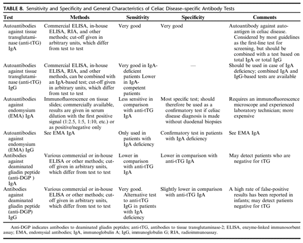
The WGO notes two main groups of serological markers for untreated CD:
- Autoantibodies targeting the auto-antigen: EMA and anti-tTG antibodies
- Antibodies targeting the offending agent (gliadin): anti-bodies against synthetic deamidated gliadin peptides (anti-DGPs)
A summary of the characteristics of CD antibody tests is listed below:
The WGO also lists several conditions associated with a higher risk of CD. Those conditions are as follows:
- Type 1 diabetes mellitus
- Autoimmune thyroid disease
- Autoimmune liver disease
- Down syndrome
- Turner syndrome
- Williams syndrome
- Selective IgA deficiency
- Unexplained elevated serum aminotransferase levels
The WGO also recommends that first-degree relatives of index (affected) patients to be screened for CD.
Finally, WGO recommends against use of urine, stool, or saliva measurements in clinical practice, as they have a “lower performance” than blood-based tests.35
European Society for the Study of Coeliac Disease (ESsCD)
The ESsCD published guidelines on CD, including recommendations on serological and genetic testing. These recommendations are listed below:
- “Adult patients with symptoms, signs or laboratory evidence suggestive of malabsorption should be tested with serology for CD. (Strong recommendation, high level of evidence)”
- “Screening of asymptomatic first-degree family member of CD patient is recommended. If available, HLA-typing may be offered as the first-line test; if negative, no further work-up is needed. (Conditional recommendation, high level of evidence)”
- “CD should be excluded in patients with unexplained elevation of serum aminotransferase levels. (Strong recommendation, high level of evidence)”
- “T1DM should be screened regularly for CD. (Strong recommendation, high level of evidence)”
- “IgA-TG2 antibody is the preferred single test for detection of CD at any age. (Strong recommendation, high level of evidence)”
- “Total IgA level needs to be measured concurrently with serology testing to determine whether IgA levels are sufficient. (Strong recommendation, moderate level of evidence)”
- “In patients with selective total IgA-deficiency, IgG-based testing (IgG-DGPs or IgG-TG2) should be performed at diagnosis and follow-up. (Strong recommendation, moderate level of evidence)”
- “All diagnostic serologic testing should be done while patients on a gluten-containing diet. (Strong recommendation, high level of evidence)”
- “Antibodies directed against native gliadin (AGA) are not recommended for the primary detection of CD. (Strong recommendation, high level of evidence)”
- “Intestinal-permeability tests are neither sensitive nor specific and are not recommended for CD diagnosis. (Strong recommendation, moderate level of evidence)”
- “Serum I-FABP might be useful in identifying dietary non-adherence and unintentional gluten intake. (Strong recommendation, moderate level of evidence)”
- “A newly diagnosed adult CD patient should undergo testing to uncover deficiencies of essential micronutrient, e.g. iron, folic acid, vitamin D and vitamin B12. (Strong recommendation, moderate level of evidence)”
- “CD diagnosis may be made without duodenal biopsy in symptomatic children with high TG2 levels (>10 times ULN) and EMA in the presence of HLA-DQ2/8. The diagnosis is confirmed by an antibody decline and preferably a clinical response to a GFD”. (Conditional recommendation, moderate level of evidence)
The ESsCD also lists recommendations for HLA-DQ2/8 typing, which are as follows:
- “A negative HLA test is helpful to exclude the possibility of CD. This is especially helpful in those already on a GFD before testing.”
- “When diagnosis of CD is uncertain, e.g., negative serology, but histology suggestive of CD.”
- “To distinguish siblings who can be reassured that it is unlikely that they will develop CD from those who need to be monitored. Furthermore, the data on the quality of life on a GFD in those patients detected by screening are conflicting, but there is a trend towards improvement. Also, the lack of understanding of the natural history of undiagnosed CD may justify screening asymptomatic persons.”
- “In subjects with other autoimmune diseases and some genetic disorders who should be investigated for CD.”
- “HLA-DQ2/DQ8 testing should not be used routinely in the initial diagnosis of CD. It is recommended that the results of such testing should be included along with a caution that patients at risk should be serologically tested for CD without changing their diet. (Strong recommendation, moderate level of evidence).”36
British Society of Gastroenterology (BSG)
In 2014, the BSG published guidelines for the diagnosis and management of adult CD. The following guidelines were included:
- “Diagnosis of CD requires duodenal biopsy when the patient is on a gluten-containing diet and for the vast majority of adult patients also positive serology. (Grade B)
- Biopsy remains essential for the diagnosis of adult CD and cannot be replaced by serology. Follow-up should aim at strict adherence to a gluten-free diet. (Grade B)
- In individuals undergoing an upper endoscopy in whom laboratory tests or symptoms or endoscopic features suggest CD, duodenal biopsy should be considered. (Grade C)
- HLA typing should be used to rule out CD. A positive DQ2.5 or DQ8 can never confirm the diagnosis. (Grade B)
- HLA typing should be used in individuals who are self-treated on a GFD and never had appropriate testing for CD before changing their diet. (Grade B)
- HLA typing can be used to rule out CD, and minimise future testing, in high-risk individuals with CD, for example, first-degree relatives. (Grade B)
- The diagnosis of CD requires duodenal biopsy when the patient is on a gluten-containing diet and for the vast majority of adult patients also positive serology. (Grade B)
- Duodenal biopsy should be retained as the mainstay for the diagnosis of adult CD and cannot be replaced by serology. (Grade B)
- At endoscopy, if there is suspicion of CD, then at least four biopsy specimens should be obtained, including a duodenal bulb biopsy. (Grade C)
- In serologically negative patients showing signs of malabsorption (such as anaemia or diarrhoea) or a family history of CD, a duodenal biopsy should be considered. (Grade C)
- Follow-up biopsies may be considered in patients with CD, and are potentially helpful in identifying patients at increased risk of lymphoma. (Grade B)”37
American Academy of Family Physicians (AAFP)
In January 2014, the AAFP released a set of recommendations regarding the diagnosis and management of celiac disease. Based on “consensus, disease-oriented evidence, usual practice, expert opinion, or case series (Evidence Rating: C),” the AAFP stated the following:
- “Immunoglobulin A tissue transglutaminase should be used as the first-line test for serologic diagnosis of suspected celiac disease.
- Small bowel biopsy should be used to confirm the diagnosis of celiac disease in most patients.”38
A 2017 the AAFP adduces the USPSTF guidelines in their recommendation statement for the screening of celiac disease. The table the AAFP included from the USPSTF is shown below.
References:
1. CDF. What is Celiac disease? Celiac Disease Foundation. https://celiac.org/celiac-disease/understanding-celiac-disease-2/what-is-celiac-disease/
2. Husby S, Koletzko S, Korponay-Szabó I, et al. European Society Paediatric Gastroenterology, Hepatology and Nutrition Guidelines for Diagnosing Coeliac Disease 2020. J Pediatr Gastroenterol Nutr. Jan 2020;70(1):141-156. doi:10.1097/mpg.0000000000002497
3. NICE. Coeliac disease overview. https://pathways.nice.org.uk/pathways/coeliac-disease
4. NIDDK. Symptoms & Causes of Celiac Disease. U.S. Department of Health and Human Services. Updated October 2020. https://www.niddk.nih.gov/health-information/digestive-diseases/celiac-disease/symptoms-causes
5. Kelly CP. Diagnosis of celiac disease in adults. Updated March 14, 2025. https://www.uptodate.com/contents/diagnosis-of-celiac-disease-in-adults
6. Nellikkal SS, Hafed Y, Larson JJ, Murray JA, Absah I. High Prevalence of Celiac Disease Among Screened First-Degree Relatives. Mayo Clin Proc. Sep 2019;94(9):1807-1813. doi:10.1016/j.mayocp.2019.03.027
7. Caio G, Volta U, Sapone A, et al. Celiac disease: a comprehensive current review. BMC Med. Jul 23 2019;17(1):142. doi:10.1186/s12916-019-1380-z
8. NIDDK. Definition & Facts for Celiac Disease. National Institute of Diabetes and Digestive and Kidney Diseases. Updated October 2020. https://www.niddk.nih.gov/health-information/digestive-diseases/celiac-disease/definition-facts
9. Brown NK, Guandalini S, Semrad C, Kupfer SS. A Clinician's Guide to Celiac Disease HLA Genetics. Am J Gastroenterol. Oct 2019;114(10):1587-1592. doi:10.14309/ajg.0000000000000310
10. Tye-Din JA, Galipeau HJ, Agardh D. Celiac Disease: A Review of Current Concepts in Pathogenesis, Prevention, and Novel Therapies. Frontiers in pediatrics. 2018;6:350. doi:10.3389/fped.2018.00350
11. Stankovic B, Radlovic N, Lekovic Z, et al. HLA genotyping in pediatric celiac disease patients. Bosnian journal of basic medical sciences. Aug 16 2014;14(3):171-6. doi:10.17305/bjbms.2014.3.28
12. Paul SP, Hoghton M, Sandhu B. Limited role of HLA DQ2/8 genotyping in diagnosing coeliac disease. Scottish medical journal. Feb 2017;62(1):25-27. doi:10.1177/0036933016689008
13. Sarna VK, Lundin KEA, Morkrid L, Qiao SW, Sollid LM, Christophersen A. HLA-DQ-Gluten Tetramer Blood Test Accurately Identifies Patients With and Without Celiac Disease in Absence of Gluten Consumption. Gastroenterology. Mar 2018;154(4):886-896.e6. doi:10.1053/j.gastro.2017.11.006
14. Lau MS, Mooney P, White W, et al. Pre-endoscopy point of care test (Simtomax- IgA/IgG-Deamidated Gliadin Peptide) for coeliac disease in iron deficiency anaemia: diagnostic accuracy and a cost saving economic model. BMC Gastroenterol. Sep 15 2016;16:115. doi:10.1186/s12876-016-0521-5
15. FDA. Decision Summary. https://www.accessdata.fda.gov/cdrh_docs/reviews/DEN160026.pdf
16. Olen O, Gudjonsdottir AH, Browaldh L, et al. Antibodies against deamidated gliadin peptides and tissue transglutaminase for diagnosis of pediatric celiac disease. J Pediatr Gastroenterol Nutr. Dec 2012;55(6):695-700. doi:10.1097/MPG.0b013e3182645c54
17. Sakly W, Mankai A, Ghdess A, Achour A, Thabet Y, Ghedira I. Performance of anti-deamidated gliadin peptides antibodies in celiac disease diagnosis. Clinics and research in hepatology and gastroenterology. Dec 2012;36(6):598-603. doi:10.1016/j.clinre.2012.01.008
18. Bufler P, Heilig G, Ossiander G, Freudenberg F, Grote V, Koletzko S. Diagnostic performance of three serologic tests in childhood celiac disease. Zeitschrift fur Gastroenterologie. Feb 2015;53(2):108-14. doi:10.1055/s-0034-1385704
19. Silvester JA, Kurada S, Szwajcer A, Kelly CP, Leffler DA, Duerksen DR. Tests for Serum Transglutaminase and Endomysial Antibodies Do Not Detect Most Patients With Celiac Disease and Persistent Villous Atrophy on Gluten-free Diets: a Meta-analysis. Gastroenterology. Sep 2017;153(3):689-701.e1. doi:10.1053/j.gastro.2017.05.015
20. Selleski N, Almeida LM, Almeida FC, Pratesi CB, Nobrega YKM, Gandolfi L. PREVALENCE OF CELIAC DISEASE PREDISPOSING GENOTYPES, INCLUDING HLA-DQ2.2 VARIANT, IN BRAZILIAN CHILDREN. Arquivos de gastroenterologia. Jan-Mar 2018;55(1):82-85. doi:10.1590/s0004-2803.201800000-16
21. Mubarak A, Spierings E, Wolters V, van Hoogstraten I, Kneepkens CM, Houwen R. Human leukocyte antigen DQ2.2 and celiac disease. J Pediatr Gastroenterol Nutr. Apr 2013;56(4):428-30. doi:10.1097/MPG.0b013e31827913f9
22. Vijzelaar R, van der Zwan E, van Gammeren A, et al. Rapid Detection of the Three Celiac Disease Risk Genotypes HLA-DQ2.2, HLA-DQ2.5, and HLA-DQ8 by Multiplex Ligation-Dependent Probe Amplification. Genetic testing and molecular biomarkers. Mar 2016;20(3):158-61. doi:10.1089/gtmb.2015.0233
23. Bajor J, Szakács Z, Farkas N, et al. Classical celiac disease is more frequent with a double dose of HLA-DQB1*02: A systematic review with meta-analysis. PloS one. 2019;14(2):e0212329. doi:10.1371/journal.pone.0212329
24. Tangermann P, Branchi F, Itzlinger A, et al. Low Sensitivity of Simtomax Point of Care Test in Detection of Celiac Disease in a Prospective Multicenter Study. Clin Gastroenterol Hepatol. Aug 2019;17(9):1780-1787.e5. doi:10.1016/j.cgh.2018.09.032
25. Profaizer T, Pole A, Monds C, Delgado JC, Lázár-Molnár E. Clinical utility of next generation sequencing based HLA typing for disease association and pharmacogenetic testing. Hum Immunol. Jul 2020;81(7):354-360. doi:10.1016/j.humimm.2020.05.001
26. Gould MJ, Mahmud FH, Clarke ABM, et al. Accuracy of Screening Tests for Celiac Disease in Asymptomatic Patients With Type 1 Diabetes. Am J Gastroenterol. Jul 1 2021;116(7):1545-1549. doi:10.14309/ajg.0000000000001193
27. Rubio-Tapia A, Hill ID, Semrad C, et al. American College of Gastroenterology Guidelines Update: Diagnosis and Management of Celiac Disease. Am J Gastroenterol. Jan 1 2023;118(1):59-76. doi:10.14309/ajg.0000000000002075
28. Husby S, Murray JA, Katzka DA. AGA Clinical Practice Update on Diagnosis and Monitoring of Celiac Disease - Changing Utility of Serology and Histologic Measures: Expert Review. Gastroenterology. 2019;156(4):885-889. doi:10.1053/j.gastro.2018.12.010
29. Green PHR, Paski S, Ko CW, Rubio-Tapia A. AGA Clinical Practice Update on Management of Refractory Celiac Disease: Expert Review. Gastroenterology. Nov 2022;163(5):1461-1469. doi:10.1053/j.gastro.2022.07.086
30. Mearin ML, Agardh D, Antunes H, et al. ESPGHAN Position Paper on Management and Follow-up of Children and Adolescents With Celiac Disease. J Pediatr Gastroenterol Nutr. Sep 1 2022;75(3):369-386. doi:10.1097/mpg.0000000000003540
31. Hill ID, Fasano A, Guandalini S, et al. NASPGHAN Clinical Report on the Diagnosis and Treatment of Gluten-related Disorders. J Pediatr Gastroenterol Nutr. Jul 2016;63(1):156-65. doi:10.1097/mpg.0000000000001216
32. NICE. Coeliac disease: recognition, assessment and management. National Institute for Health and Care Excellence. https://www.nice.org.uk/guidance/ng20/resources/coeliac-disease-recognition-assessment-and-management-pdf-1837325178565
33. NICE. Coeliac disease. National Institute for Health and Care Excellence. https://www.nice.org.uk/guidance/qs134/resources/coeliac-disease-pdf-75545419042501
34. Bibbins-Domingo K, Grossman DC, Curry SJ, et al. Screening for Celiac Disease: US Preventive Services Task Force Recommendation Statement. Jama. Mar 28 2017;317(12):1252-1257. doi:10.1001/jama.2017.1462
35. Bai JC, Ciacci C. World Gastroenterology Organisation Global Guidelines: Celiac Disease February 2017. Journal of clinical gastroenterology. Oct 2017;51(9):755-768. doi:10.1097/mcg.0000000000000919
36. Al-Toma A, Volta U, Auricchio R, et al. European Society for the Study of Coeliac Disease (ESsCD) guideline for coeliac disease and other gluten-related disorders. United European gastroenterology journal. Jun 2019;7(5):583-613. doi:10.1177/2050640619844125
37. Ludvigsson JF, Bai JC, Biagi F, et al. Diagnosis and management of adult coeliac disease: guidelines from the British Society of Gastroenterology. Gut. Aug 2014;63(8):1210-28. doi:10.1136/gutjnl-
38. Pelkowski TD, Viera AJ. Celiac disease: diagnosis and management. Am Fam Physician. Jan 15 2014;89(2):99-105.
39. AAFP. Screening for Celiac Disease: Recommendation Statement. Am Fam Physician. Sep 15 2017;96(6):Online.
40. FDA. Ig_Plex Celiac DGP Panel. https://www.accessdata.fda.gov/scripts/cdrh/devicesatfda/index.cfm?db=pmn&id=K140691
41. FDA. Aptiva Celiac Disease IgA Reagent. https://www.accessdata.fda.gov/cdrh_docs/reviews/K193604.pdf
Coding Section
| Code |
Number |
Description |
| CPT |
82784 |
Gammaglobulin (immunoglobulin); IgA, IgD, IgG, IgM, each |
|
|
83516 |
Immunoassay for analyte other than infectious agent antibody or infectious agent antigen; qualitative or semiquantitative, multiple method |
|
|
86231 (effective 01/01/2022) |
Endomysial antibody (EMA), each immunoglobulin (Ig) class |
|
|
86255 |
Fluorescent noninfectious agent anitbody, screen, each antibody |
|
|
86256 |
Titer, each antibody (fluorescent technique for antigen identification in tissue, use 88346, for indirect fluorescence, use 88347) (FTA, use 86780) (Gel [agar] diffusion tests, use 86331) |
|
|
86258 (effective 01/01/2022) |
Gliadin (deamidated) (DGP) antibody, each immunoglobulin (Ig) class |
|
|
86364 (effective 01/01/2022) |
Tissue transglutaminase, each immunoglobulin (Ig) class |
|
|
88305 |
Level IV - Surgical pathology, gross and microscopic examination, colon biopsy |
| ICD-10-CM (effective 10/01/15) |
K90.0 |
Celiac disease |
|
|
E30.0 |
Delayed puberty |
|
|
E53.8 |
Deficiency of other specified B group vitamins |
|
|
E53.9 |
Vitamin B deficiency, unspecified |
|
|
E10.10-E10.9 |
Type 1 diabetes mellitus |
|
|
M80.00XA-M80.88XS |
Age-related osteoporosis with current pathological fracture, unspecified site |
|
|
M81.6 |
Localized osteoporosis (Lequesne) |
|
|
M81.8 |
Other osteoporosis without current pathological fracture |
|
|
M83.0-M83.9 |
Adult osteomalacia |
|
|
N91.0-N91.2 |
Absent, scanty and rare menstruation |
|
|
O03.0-O03.9 |
Spontaneous abortion |
|
|
Q96.0-Q96.9 |
Turner's syndrome |
|
|
Q93.82 |
Williams syndrome |
|
|
R62.7 |
Adult failure to thrive |
|
|
R62.51 |
Failure to thrive (child) |
|
|
R62.52 |
Short stature (child) |
|
|
R11.0-R11.9 |
Nausea and vomiting |
| ICD-10-PCS (effective 10/01/15) |
|
Not applicable. ICD-10-PCS codes are only used for inpatient services. There are no ICD procedure codes for laboratory tests. |
Procedure and diagnosis codes on Medical Policy documents are included only as a general reference tool for each policy. They may not be all-inclusive.
This medical policy was developed through consideration of peer-reviewed medical literature generally recognized by the relevant medical community, U.S. FDA approval status, nationally accepted standards of medical practice and accepted standards of medical practice in this community, and other nonaffiliated technology evaluation centers, reference to federal regulations, other plan medical policies, and accredited national guidelines.
"Current Procedural Terminology © American Medical Association. All Rights Reserved"
History From 2014 Forward
| 10/14/2025 | Annual review, removing coverage criteria which referred to genetic testing. Also updating background, regulatory status, table of terminology, rationale, and references. Removed CPT 81376, 81377, 81382, 81383. |
| 10/10/2024 | Annual review, no change to policy intent. Updating table of terminology, rationale and references. |
| 10/25/2023 | Annual review, no change to policy intent. Entire policy updated for clarity and consistency. Adding new coverage criteria #1 and 2 to address TTG testing for individuals diagnosed with celiac disease who are IgA sufficient and IgA deficiency with frequency limitations provided.) |
| 11/08/2022 | Annual review, no change to policy intent. Policy reformatted for clarity. Adding table of terminology. Updating rationale, references and coding. |
| 12/8/2021 |
Updating policy with 2022 coding. Adding code 86231, 86258 and 86364. No other change made. |
| 10/01/2021 |
Annual review, no change to policy intent. Updating background, regulatory status, rationale, references and policy number. |
| 10/01/2020 |
Annual review, reformatting policy and adding "note 1" for clarity. Updating coding, description, rationale and references. |
| 10/17/2019 |
Annual review, no change to policy intent. Reformatting for clarity, updating coding. |
| 11/05/2018 |
Annual review, adding HLA-DQ- gluten testing as investigational, also adding rapid antigen point of care testing for anti-TTG as not medically necessary. Adding panel testing, multiples or multi analyte testing as not medically necessary. No other changes. |
| 11/02/2017 |
Annual review, updating medical necessity criteria for more specificity regarding symptoms suggestive of celiac disease. No change to policy |
| 04/26/2017 |
Updated category to Laboratory. No other changes. |
| 04/05/2017 |
Annual review, no change to policy intent. |
| 06/13/2016 |
Corrected a typo error in the policy section. |
| 05/09/2016 |
Interim review adding criteria in policy section. |
| 04/07/2015 |
Annual review, no change to policy intent. Adding coding. |
| 04/10/2014 |
Annual review. Adding related policies and benefit application. Updated guidelines. No change to policy intent. |